
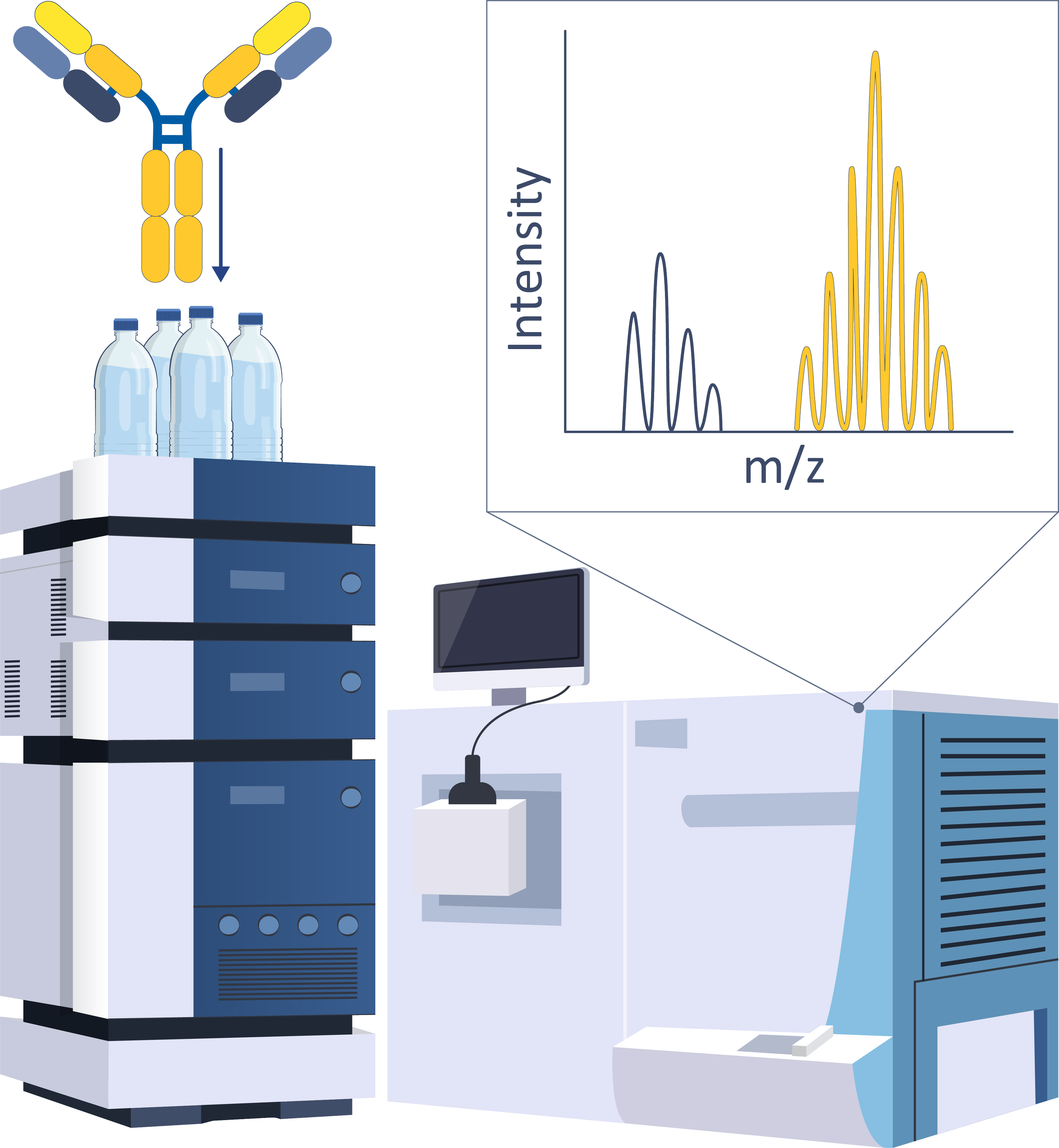
Intact Mass Spectrometry
High-resolution analysis of intact proteins to determine molecular mass and evaluate structural integrity.
Workflow:
→ Direct infusion or LC-MS
→ Deconvolution
→ Mass confirmation
Applications:
• Mass confirmation of expressed proteins
• Identification of clipping, aggregation, or degradation
• Detection of high-abundance PTMs
• Batch-to-batch comparison for biologics manufacturing

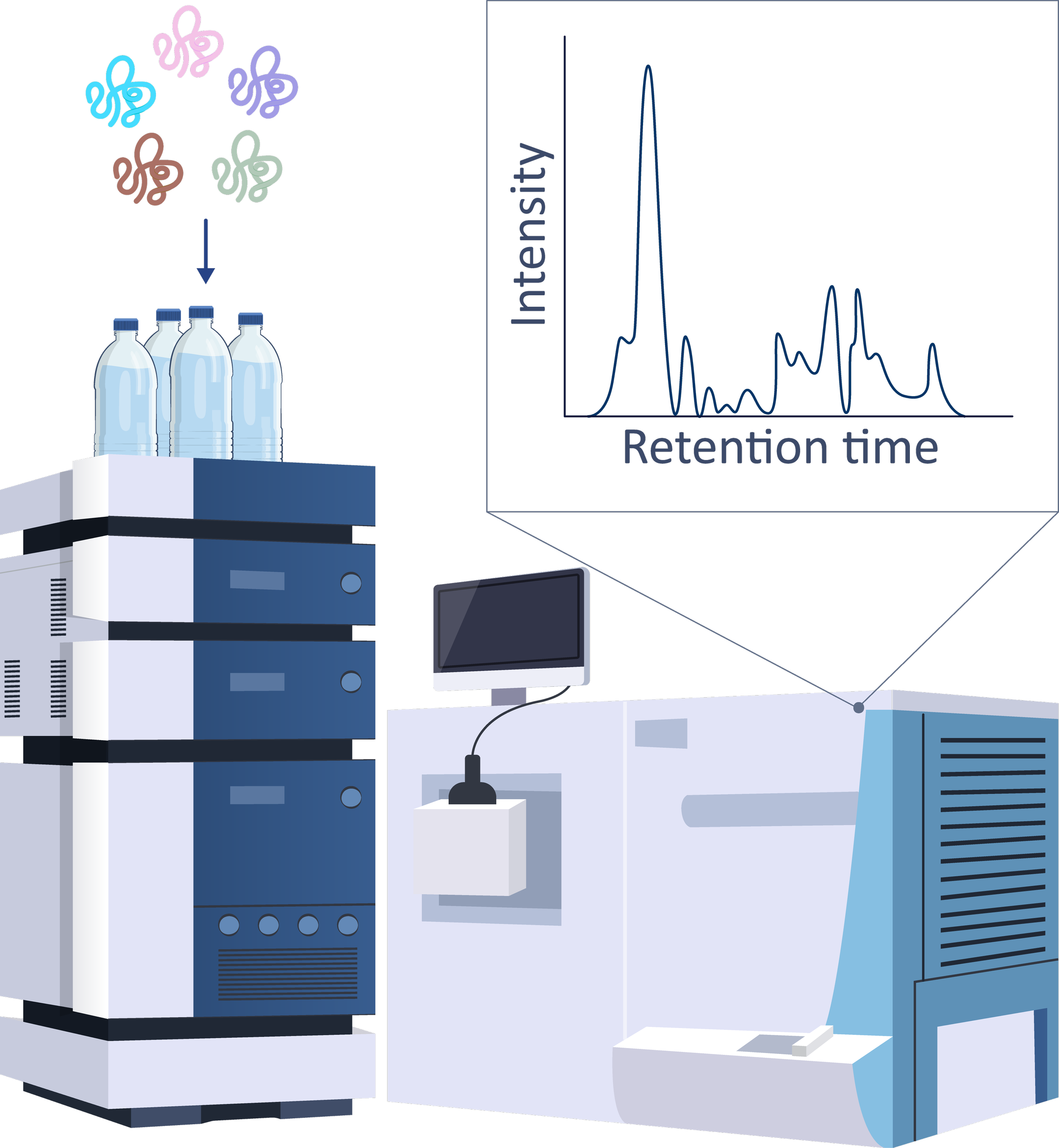
Peptide Mapping
Workflow:
→ Proteolytic digestion
→ LC-MS/MS analysis
→ Data interpretation
High-resolution peptide mapping enables confirmation of protein sequence and localization of post-translational modifications (PTMs), forming the backbone of Multi-Attribute Monitoring (MAM) workflows.
Applications:
• Sequence confirmation of recombinant proteins
• Detection and quantitation of PTMs (e.g., oxidation, deamidation, etc.)
• Support for MAM-based QC strategies
• Monitoring and validation of CQAs
• Batch comparability and lot release testing
Proteomics
Top-down and bottom-up proteomics to characterize proteins in complex samples or purified forms.
Workflow:
→ Sample prep
→ LC-MS/MS (intact or digested)
→ Database search and analysis
Applications:
• Protein identification and quantitation
• Drug target discovery and validation
• PTM analysis
• Functional and interaction profiling
• Biomanufacturing QC

Glyco-Profiling
Mass spectrometry-based identification and characterization of glycan structures on proteins.
Workflow:
→ Enzymatic or chemical release
→ MS/MS analysis
→ Glycan structure annotation
Applications:
• Glycosylation site identification
• Glycan structure and branching analysis
• Quality control in biologics
• Mechanistic studies of disease-related glycosylation
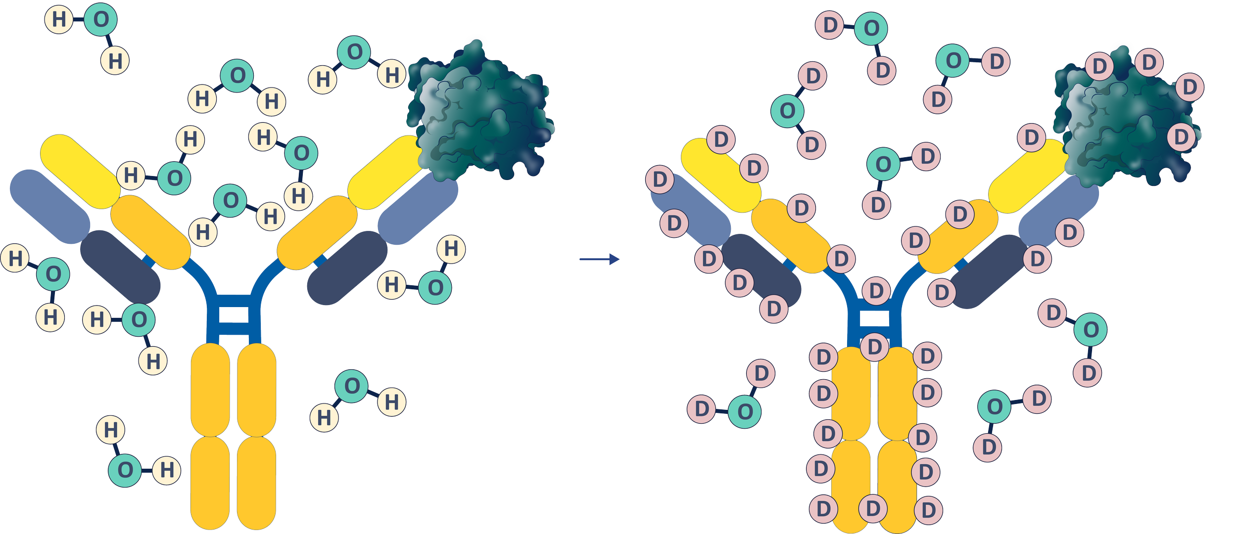
HDX-MS
Hydrogen Deuterium Exchange Mass Spectrometry reveals protein dynamics, conformational stability, and binding interfaces through deuterium uptake profiling.
Workflow:
→ Deuterium labeling
→ Quenching and digestion
→ LC-MS/MS
→ Uptake comparison
Applications:
• Epitope and paratope mapping
• Protein-ligand and protein-protein interaction studies
• Structural comparability and biosimilarity
• Biologics formulation optimization
Polyclonal Epitope Mapping
A proprietary HDX-MS-based workflow for mapping dominant epitopes within polyclonal serum in complex biological matrices.
Workflow:
→ Serum binding
→ HDX-MS analysis
→ Epitope landscape mapping
Applications:
• Vaccine response profiling
• Polyclonal antibody characterization
• Immunogenicity and serological surveillance.
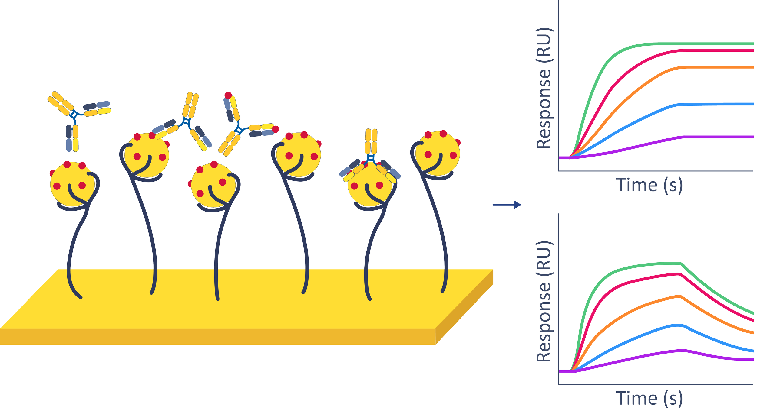
BLI
BioLayer Interferometry is a label-free optical technique that quantifies real-time biomolecular interactions, including association and dissociation kinetics.
Workflow:
→ Ligand immobilization
→ Analyte binding
→ Real-time optical readout
Applications:
• Kinetic (on/off-rate) analysis
• Affinity comparison across variants
• Screening for binding strength and specificity
